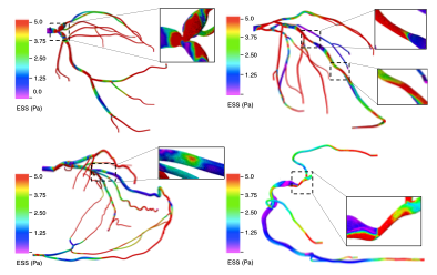
A multi-disciplinary team like by Amanda Randles at Duke University is using high-resolution patient-specific simulations to better understand coronary artery disease. Conventional invasive diagnostic imaging techniques do not adequately resolve complex coronary lesions, which present unique challenges, require personalized treatment and result in worsened patient outcomes. These lesions are often excluded from large-scale non-invasive clinical trials and there does not exist a validated approach to characterize hemodynamic quantities and guide percutaneous intervention for such lesions. This work identifies key biomarkers that differentiate complex lesions from simple lesions by introducing and validating a coronary angiography-based computational fluid dynamic framework for intracoronary assessment in complex lesions at ultrahigh resolution. Biomarkers from hemodynamic evaluation can provide much the needed insight into the increase in adverse outcomes for such patients and has incremental prognostic value over traditional pressure-based indices, such as fractional flow reserve. [Vardhan et al., “Non-invasive characterization of complex coronary lesions,” Scientific Reports 11, 2021. DOI: https://doi.org/10.1038/s41598-021-86360-6]
