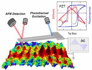
Elasticity is closely related to the chemical composition, crystal structure and mesoscopic domain configuration of materials. Contact resonance voltage spectroscopy on an atomic force microscope allows, for the first time, nanoscale elastic moduli and loss factors to be quantitatively tracked during tip bias-induced transitions. Local polarization switching processes in true ferroelectric materials are accompanied by characteristic changes in both stiffness and loss factor due to domain structure evolution, which are absent in charge-injection phenomena. This may offer an approach to identify unknown ferroelectric materials. Contact resonance voltage spectroscopy may find applications in a broad spectrum of nanoscience, for example, battery materials where Li-ion (de-)intercalation usually induces significant stiffness changes in the materials.
Q. Li, S. Jesse, A. Tselev, L. Collins, P. Yu, I. I. Kravchenko, S. V. Kalinin, and N. Balke, “Probing Local Bias-Induced Transitions Using Photothermal Excitation Contact Resonance Atomic Force Microscopy and Voltage Spectroscopy,” ACS Nano 9, 1848 (2015). DOI:10.1021/nn506753u
For more information

