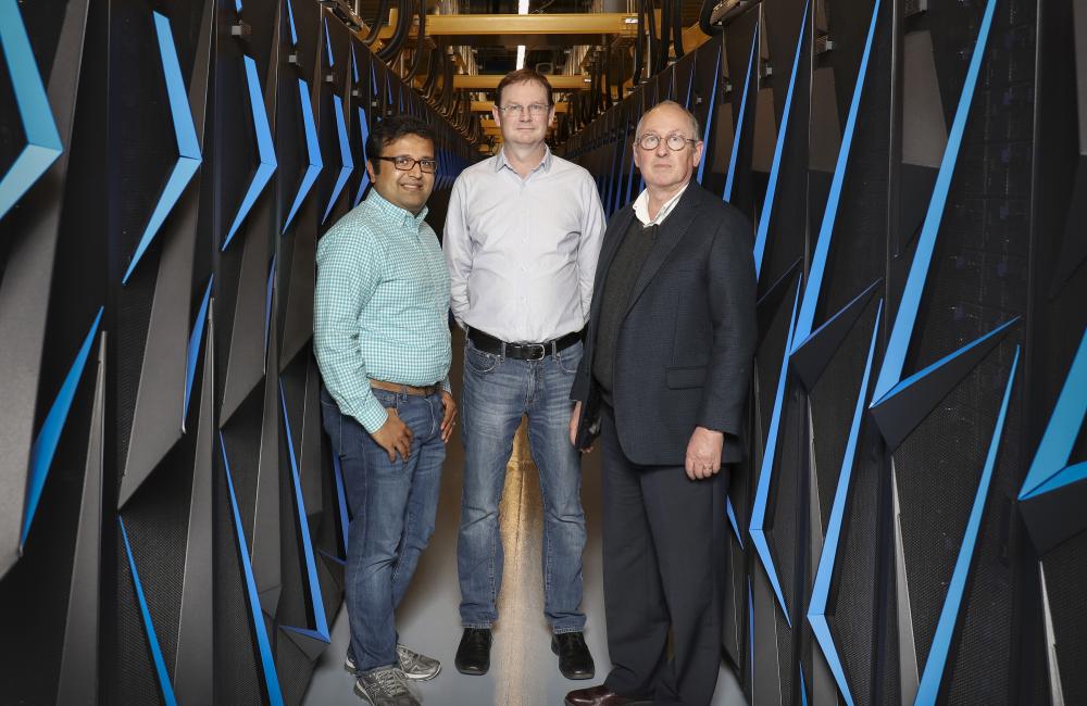
April 18, 2018—Nearly a dozen scientists across Oak Ridge National Laboratory are teaming with medical researchers and leveraging ORNL’s biggest science tools to solve a modern-day biology grand challenge: unlocking the secrets of disordered proteins. These flexible molecules are believed to constitute as much as half of the proteins in the human body yet are poorly understood because we have not found a way to adequately study their properties.
It is only in the last decade that scientists have come to accept that anywhere from one-third to one-half of human proteins do not follow the once-sacrosanct rule of molecular biology: proteins fold into stable, three-dimensional shapes. Instead, disordered proteins are constantly cycling between different forms. They are essential to cell circuitry, and their misfunction is directly implicated in diseases such as cancer, Alzheimer’s, cardiovascular conditions, and diabetes. Understanding their complex nature could lead to important new drug discoveries.
In a lab-directed research project launched this year, ORNL scientists are combining experimentation and simulation in an effort to bring clarity to the inner workings of these proteins. The project includes collaborators from the Frederick National Laboratory for Cancer Research—sponsored by the National Cancer Institute as part of the National Institutes of Health (NIH).
The internal motions of disordered proteins make them particularly hard to characterize, noted Arvind Ramanathan of the Computational Science and Engineering Division and the Health Data Sciences Institute at ORNL. The proteins defy the standard tools for characterization such as x-ray crystallography because they resist being crystallized.
“It’s like taking 2D photos of someone from different orientations and suddenly you’re asked to make a 3D rendering of that person,” Ramanathan said.
“Now imagine if that person is jumping around. You’re going to get lots of weird features in that rendering and some parts of it may even vanish,” said principal investigator Hugh O’Neill of the Neutron Scattering Division.
The researchers will use neutron scattering at the Spallation Neutron Source at ORNL and cryo-electron microscopy (cryo-EM) images from the Frederick National Laboratory to give a good estimate of what the particles look like in terms of overall shape and size, and to supply reference points for a 3D model.
“We are excited to see more and more laboratories taking advantage of our shared cryo-EM facility,” said Ethan Dmitrovsky, M.D., president of Leidos Biomedical Research, Inc. and laboratory director of the Frederick National Laboratory. “This particular project has special potential for a new area of research that might ease the suffering of patients with cancer and other illnesses.”
Neutrons are sensitive to hydrogen, non-destructive, and they make it possible to study the proteins in real time, under real-world conditions. The cryo-EM method carried out at Frederick images frozen, hydrated specimens, allowing molecular resolution without the need for dyes or fixatives. Researchers will specifically target the neurofibromatosis type 1 protein and its interactions with binding partners. Mutations in NF1 are known to cause neurofibromatosis and have been implicated in cancer.
The data will go through a process of algorithm-guided reconstruction to eliminate “noise” in the images. Then the resulting model will be used in machine learning-aided computer simulations to explore certain regions of the proteins to gain a better understanding of particle orientation.
Combining cryo-EM, small angle scattering, and computation will make it possible to generate atomistic models to reach sub-nanometer resolution for these proteins.
“Basically, the experimentalists will handle the small angle scattering, the crystallography work and the cryo-EM. Then the computer scientists will take all this disparate experimental data and put that together to give us a picture of what the protein looks like,” O’Neill said.
The project involves scientists from three directorates at ORNL: Neutron Sciences, Computing and Computational Sciences, and Energy and Environmental Sciences.
“Computation ties it all together. That will become, I think, very common in structural biology—this idea of integrating different experimental modalities that are tied together by computation,” Ramanathan added.
In fact, the scientists expect that the computational work will be among the first projects to utilize Summit, slated to come online this year as the world’s smartest, open-source supercomputer for artificial intelligence applications at DOE’s Oak Ridge Leadership Computing Facility (OLCF) at ORNL.
“This work serves as a wonderful example of how we can combine multiple science user facilities, in this case, the OLCF, the [DOE] Spallation Neutron Source, and Frederick National Laboratory’s National Cryo-Electron Microscopy Facility at Frederick National Laboratory to advance both DOE and NIH missions,” said Paul Gilna, director of biosecurity and biomedical initiatives at ORNL.
The research has applications not only for ORNL’s work in the biomedical space but is also pertinent to its work on bioenergy and mercury toxicity—areas relevant to DOE’s Biological and Environmental Research program. The research could, for instance, help scientists engineer microbes that are better at digesting and converting feedstock plants into biofuels.
The Frederick National Laboratory for Cancer Research is dedicated to improving human health through discovery and innovation in the biomedical sciences, focusing on cancer, AIDS, and rapid response to emerging infectious diseases. The Frederick National Laboratory is operated by Leidos Biomedical Research, Inc. under contract with the National Cancer Institute. SNS and OLCF are DOE Office of Science user facilities.
ORNL is managed by UT-Battelle for the Department of Energy's Office of Science, the single largest supporter of basic research in the physical sciences in the United States. DOE’s Office of Science is working to address some of the most pressing challenges of our time. For more information, please visit http://energy.gov/science.


