PI: Yanwen Zhang
By correlating electron microscopy and atom probe tomography (APT) with advanced simulations, researchers revealed irradiation-induced chemical segregation and laid the foundation for APT imaging of nanoscale voids. Understanding the imaging process enables chemical composition measurement near voids with unprecedented accuracy in various materials.
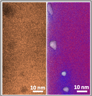
Void formation is a major reason for nuclear materials degradation. An accurate measurement of radiation-induced segregation around voids is critical for understanding and suppressing materials degradation. APT is up to the challenge of improving accuracy because it can reconstruct element positions in three dimensions with high spatial resolution and chemical sensitivity. However, a knowledge gap exists regarding how APT images voids. By analyzing the same voids using both electron microscopy and APT, researchers demonstrated that in APT data, local atomic densities near voids can differ. Simulations revealed the physical mechanisms for these density changes and how to interpret them. This work reveals chemically biased mass transport in next generation alloys under irradiation and provides a general approach for using APT to quantify material chemistries near voids with 10 parts per million element sensitivities.
Related Publication:
X. Wang, C. Hatzoglou, B. Sneed, Z. Fan, W. Guo, K. Jin, D. Chen, H. Bei, Y. Wang, W. J. Weber, Y. Zhang, B. Gault, K. L. More, F. Vurpillot, and J. D. Poplawsky, Interpreting nano-voids in atom probe tomography for accurate local composition measurements, Nature Commun. 11, 1022 (2020).