![Sphere that has the top right fourth removed (exposed) Colors from left are orange, dark blue with orange dots, light blue with horizontal lines, then black. Inside the exposure is green and black with boxes.](/sites/default/files/styles/featured_square_large/public/2024-06/slicer.jpg?h=56311bf6&itok=bCZz09pJ)
Filter News
Area of Research
News Topics
- (-) Artificial Intelligence (7)
- (-) Clean Water (2)
- (-) Energy Storage (6)
- (-) Microscopy (9)
- 3-D Printing/Advanced Manufacturing (7)
- Advanced Reactors (2)
- Big Data (4)
- Bioenergy (4)
- Biomedical (5)
- Composites (3)
- Computer Science (30)
- Critical Materials (1)
- Cybersecurity (2)
- Environment (6)
- Exascale Computing (2)
- Frontier (2)
- Fusion (3)
- Grid (2)
- Isotopes (6)
- Materials Science (25)
- Molten Salt (1)
- Nanotechnology (12)
- Neutron Science (8)
- Nuclear Energy (10)
- Physics (8)
- Polymers (6)
- Quantum Science (6)
- Security (1)
- Space Exploration (3)
- Summit (11)
- Sustainable Energy (6)
- Transportation (8)
Media Contacts
![ORNL researcher Karren More has been elected fellow of the Microscopy Society of America.](/sites/default/files/styles/list_page_thumbnail/public/2019-03/K_More_th.jpg?h=655057a4&itok=53tPHa-r)
OAK RIDGE, Tenn., March 22, 2019 – Karren Leslie More, a researcher at the Department of Energy’s Oak Ridge National Laboratory, has been elected fellow of the Microscopy Society of America (MSA) professional organization.
![In this MXene electrode, choosing the appropriate solvent for the electrolyte can increase energy density significantly. This scanning electron microscopy image shows fine features of a film only 5 microns thick—approximately 10 times narrower than a human hair. Credit: Drexel University; image by Tyler Mathis](/sites/default/files/styles/list_page_thumbnail/public/2019-03/MXene%20electrode_0.jpg?h=e9daaebf&itok=YNpINGl2)
![(From left) ORNL Associate Laboratory Director for Computing and Computational Sciences Jeff Nichols; ORNL Health Data Sciences Institute Director Gina Tourassi; DOE Deputy Under Secretary for Science Thomas Cubbage; ORNL Task Lead for Biostatistics Blair Christian; and ORNL Research Scientist Ioana Danciu were invited to the White House to showcase an ORNL-developed digital tool aimed at better matching cancer patients with clinical trials.](/sites/default/files/styles/list_page_thumbnail/public/2019-03/TourassiWH%5B1%5D.png?h=26b5064d&itok=HUC2iYmE)
OAK RIDGE, Tenn., March 4, 2019—A team of researchers from the Department of Energy’s Oak Ridge National Laboratory Health Data Sciences Institute have harnessed the power of artificial intelligence to better match cancer patients with clinical trials.
![To develop complex materials with superior properties, Vera Bocharova uses diverse methods including broadband dielectric spectroscopy. Credit: Oak Ridge National Laboratory, U.S. Dept. of Energy; photographer Jason Richards](/sites/default/files/styles/list_page_thumbnail/public/2019-02/2016-p05202.jpg?h=b6236d98&itok=w-Sd8giq)
Vera Bocharova at the Department of Energy’s Oak Ridge National Laboratory investigates the structure and dynamics of soft materials—polymer nanocomposites, polymer electrolytes and biological macromolecules—to advance materials and technologies for energy, medicine and other applications.
![ORNL alanine_graphic.jpg ORNL alanine_graphic.jpg](/sites/default/files/styles/list_page_thumbnail/public/ORNL%20alanine_graphic.jpg?itok=iRLfcOw-)
OAK RIDGE, Tenn., Jan. 31, 2019—A new electron microscopy technique that detects the subtle changes in the weight of proteins at the nanoscale—while keeping the sample intact—could open a new pathway for deeper, more comprehensive studies of the basic building blocks of life.
![Supercomputing-Memory_boost1.jpg Supercomputing-Memory_boost1.jpg](/sites/default/files/styles/list_page_thumbnail/public/Supercomputing-Memory_boost1.jpg?itok=dDR8CnYC)
Scientists at Oak Ridge National Laboratory and Hypres, a digital superconductor company, have tested a novel cryogenic, or low-temperature, memory cell circuit design that may boost memory storage while using less energy in future exascale and quantum computing applications.
![Picture2.png Picture2.png](/sites/default/files/styles/list_page_thumbnail/public/Picture2_1.png?itok=IV4n9XEh)
Oak Ridge National Laboratory scientists studying fuel cells as a potential alternative to internal combustion engines used sophisticated electron microscopy to investigate the benefits of replacing high-cost platinum with a lower cost, carbon-nitrogen-manganese-based catalyst.
![B_Hudak_ORNL.jpg B_Hudak_ORNL.jpg](/sites/default/files/styles/list_page_thumbnail/public/B_Hudak_ORNL.jpg?itok=Os5uKm-q)
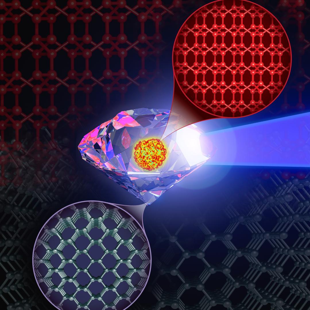
An Oak Ridge National Laboratory-led team used a scanning transmission electron microscope to selectively position single atoms below a crystal’s surface for the first time.
![Sergei Kalinin, director of the Institute for Functional Imaging of Materials at Oak Ridge National Laboratory, convenes experts in microscopy and computing to gain scientific insights that will inform design of advanced materials for energy and informati Sergei Kalinin, director of the Institute for Functional Imaging of Materials at Oak Ridge National Laboratory, convenes experts in microscopy and computing to gain scientific insights that will inform design of advanced materials for energy and informati](/sites/default/files/styles/list_page_thumbnail/public/news/images/2018-P00854%20%28002%29.jpg?itok=UfhMWf3G)
Sergei Kalinin of the Department of Energy’s Oak Ridge National Laboratory knows that seeing something is not the same as understanding it. As director of ORNL’s Institute for Functional Imaging of Materials, he convenes experts in microscopy and computing to gain scientific insigh...
![Schematic drawing of the boron nitride cell. Credit: University of Illinois at Chicago. Schematic drawing of the boron nitride cell. Credit: University of Illinois at Chicago.](/sites/default/files/styles/list_page_thumbnail/public/news/images/schematic1.jpg?itok=iYCttAg3)
A new microscopy technique developed at the University of Illinois at Chicago allows researchers to visualize liquids at the nanoscale level — about 10 times more resolution than with traditional transmission electron microscopy — for the first time. By trapping minute amounts of...