An AI-generated image representing atoms and artificial neural networks. Credit: Maxim Ziatdinov, ORNL

Researchers at ORNL have developed a machine-learning inspired software package that provides end-to-end image analysis of electron and scanning probe microscopy images.

Artificial intelligence is becoming an increasingly valuable tool for ORNL researchers tackling the many mysteries of cancer. Credit: Getty Images.

A team of researchers from ORNL was recognized by the National Cancer Institute in March for their unique contributions in the fight against cancer.

Computational systems biologists at ORNL worked with the U.S. Department of Veterans Affairs and other institutions to identify 139 locations across the human genome tied to risk factors for varicose veins, marking a first step in the development of new treatments. Credit: Andy Sproles/ORNL, U.S. Dept. of Energy

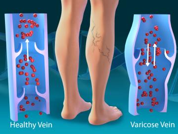
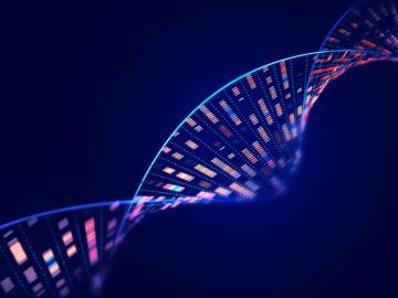
As part of a multi-institutional research project, scientists at ORNL leveraged their computational systems biology expertise and the largest, most diverse set of health data to date to explore the genetic basis of varicose veins.

When an electron beam drills holes in heated graphene, single-atom vacancies, shown in purple, diffuse until they join with other vacancies to form stationary structures and chains, shown in blue. Credit: Ondrej Dyck/ORNL, U.S. Dept. of Energy

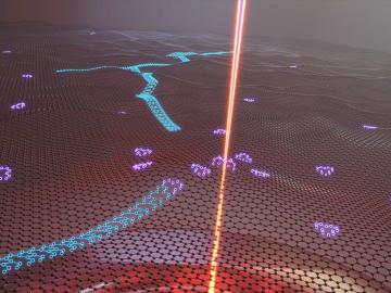
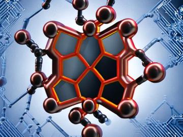
Oak Ridge National Laboratory researchers serendipitously discovered when they automated the beam of an electron microscope to precisely drill holes in the atomically thin lattice of graphene, the drilled holes closed up.

Jack Cahill of ORNL’s Biosciences Division is developing new techniques to view and measure the previously unseen to better understand important chemical processes at play in plant-microbe interactions and in human health. In this photo, Cahill is positioning a rhizosphere-on-a-chip platform for imaging by mass spectrometry. Credit: Carlos Jones/ORNL, U.S. Dept of Energy

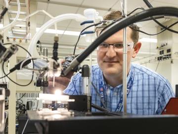
John “Jack” Cahill is out to illuminate previously unseen processes with new technology, advancing our understanding of how chemicals interact to influence complex systems whether it’s in the human body or in the world beneath our feet.

Shown here is the structure of the NEMO protein. A team from ORNL conducted extensive molecular dynamics work on Summit by using both quantum mechanics and machine-learning methods to look at the binding affinity of NEMO and 3CLpro in humans and other species and to consider the structural models derived from the sequences of other coronaviruses. Image courtesy Nature Communications, Dan Jacobson/ORNL.

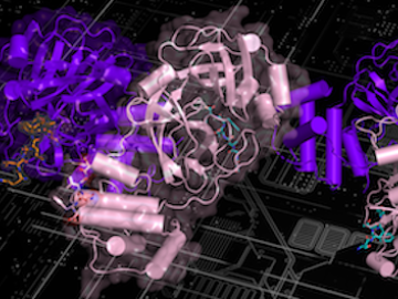
A new paper published in Nature Communications adds further evidence to the bradykinin storm theory of COVID-19’s viral pathogenesis — a theory that was posited two years ago by a team of researchers at the Department of Energy’s Oak Ridge National Laboratory.

Scientists at ORNL have created a rhizosphere-on-a-chip research platform, a miniaturized environment to study the ecosystem around poplar tree roots for insights into plant health and soil carbon sequestration. Credit: Carlos Jones/ORNL, U.S. Dept. of Energy

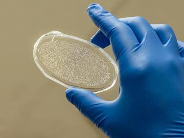
Scientists at ORNL have created a miniaturized environment to study the ecosystem around poplar tree roots for insights into plant health and soil carbon sequestration.

Larry Allard

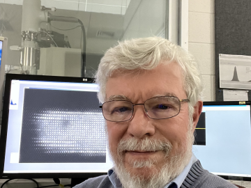
Larry Allard, a distinguished research staff member at Oak Ridge National Laboratory, has been named a Fellow of the Microanalysis Society.

Samarthya Bhagia examines a sample of a thermoplastic composite material additively manufactured using poplar wood and polylactic acid. Credit: Carlos Jones/ORNL, U.S. Dept. of Energy

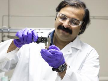
Chemical and environmental engineer Samarthya Bhagia is focused on achieving carbon neutrality and a circular economy by designing new plant-based materials for a range of applications from energy storage devices and sensors to environmentally friendly bioplastics.

Scattering-type scanning near-field optical microscopy, a nondestructive technique in which the tip of the probe of a microscope scatters pulses of light to generate a picture of a sample, allowed the team to obtain insights into the composition of plant cell walls. Credit: Ali Passian/ORNL, U.S. Dept. of Energy

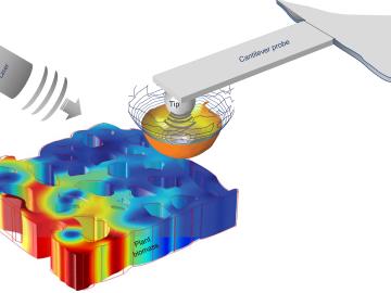
To optimize biomaterials for reliable, cost-effective paper production, building construction, and biofuel development, researchers often study the structure of plant cells using techniques such as freezing plant samples or placing them in a vacuum.