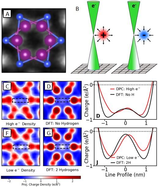
Figure: (A) Atomic structure of interstitial column in Y5Si3. (B) Schematic of DPC charge-density measurement. (C-E) Comparison of DPC in high e- sites to theoretical anionic column density. (F-H) Comparison of DPC in low e- columns to theoretical hydrogenated anionic column density.
Scientific Achievement
Anionic electrons in Y5Si3 are visualized with sub-Å spatial resolution in a scanning transmission electron microscope (STEM), revealing an unexpected charge variation at different anionic sites for the first time.
Significance and Impact
Visualizing and quantifying charge combined with theory presents new opportunities to exploit real-space charge distributions in quantum materials.
Research Details
- Differential phase contrast (DPC) imaging on a pixelated detector relates small shifts in the diffracted beam to charge-density with sub-Å spatial resolution.
- An unexpected charge variation is revealed at different anionic sites, the origin of which was trace interstitial hydrogen as confirmed by theoretical calculations and neutron spectroscopy.
Q. Zheng, T. Feng, J. A. Hachtel, R. Ishikawa, Y. Cheng, L. Daemen, J. Xing, J. C. Idrobo, J. Yan, N. Shibata, Y. Ikuhara, B. C. Sales, S. T. Panetiledes, and M. Chi, “Direct Visualization of Anionic Electrons in an Electride Reveals Inhomogeneities,” Sci. Adv. 7, eabe6819 (2021).