Filter News
Area of Research
News Topics
- (-) Nanotechnology (60)
- 3-D Printing/Advanced Manufacturing (116)
- Advanced Reactors (32)
- Artificial Intelligence (81)
- Big Data (49)
- Bioenergy (86)
- Biology (93)
- Biomedical (56)
- Biotechnology (20)
- Buildings (54)
- Chemical Sciences (55)
- Clean Water (29)
- Climate Change (91)
- Composites (25)
- Computer Science (181)
- Coronavirus (46)
- Critical Materials (23)
- Cybersecurity (35)
- Decarbonization (71)
- Education (3)
- Element Discovery (1)
- Emergency (2)
- Energy Storage (106)
- Environment (188)
- Exascale Computing (34)
- Fossil Energy (4)
- Frontier (38)
- Fusion (51)
- Grid (59)
- High-Performance Computing (80)
- Hydropower (11)
- Irradiation (3)
- Isotopes (46)
- ITER (7)
- Machine Learning (43)
- Materials (138)
- Materials Science (130)
- Mathematics (6)
- Mercury (12)
- Microelectronics (2)
- Microscopy (50)
- Molten Salt (8)
- National Security (54)
- Net Zero (10)
- Neutron Science (127)
- Nuclear Energy (101)
- Partnerships (37)
- Physics (58)
- Polymers (31)
- Quantum Computing (28)
- Quantum Science (65)
- Renewable Energy (2)
- Security (23)
- Simulation (42)
- Software (1)
- Space Exploration (24)
- Statistics (2)
- Summit (57)
- Sustainable Energy (115)
- Transformational Challenge Reactor (7)
- Transportation (92)
Media Contacts

OAK RIDGE, Tenn., Jan. 31, 2019—A new electron microscopy technique that detects the subtle changes in the weight of proteins at the nanoscale—while keeping the sample intact—could open a new pathway for deeper, more comprehensive studies of the basic building blocks of life.
![2018-P07635 BL-6 user - Univ of Guelph-6004R_sm[2].jpg 2018-P07635 BL-6 user - Univ of Guelph-6004R_sm[2].jpg](/sites/default/files/styles/list_page_thumbnail/public/2018-P07635%20BL-6%20user%20-%20Univ%20of%20Guelph-6004R_sm%5B2%5D.jpg?itok=DUdZNt_q)
A team of scientists, led by University of Guelph professor John Dutcher, are using neutrons at ORNL’s Spallation Neutron Source to unlock the secrets of natural nanoparticles that could be used to improve medicines.
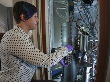
An Oak Ridge National Laboratory-led team used a scanning transmission electron microscope to selectively position single atoms below a crystal’s surface for the first time.
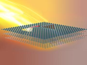
Scientists at the Department of Energy’s Oak Ridge National Laboratory induced a two-dimensional material to cannibalize itself for atomic “building blocks” from which stable structures formed. The findings, reported in Nature Communications, provide insights that ...
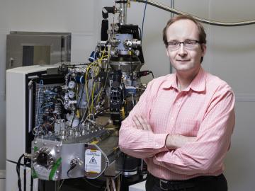
Sergei Kalinin of the Department of Energy’s Oak Ridge National Laboratory knows that seeing something is not the same as understanding it. As director of ORNL’s Institute for Functional Imaging of Materials, he convenes experts in microscopy and computing to gain scientific insigh...
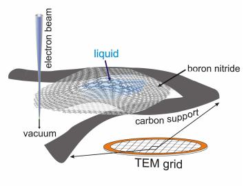
A new microscopy technique developed at the University of Illinois at Chicago allows researchers to visualize liquids at the nanoscale level — about 10 times more resolution than with traditional transmission electron microscopy — for the first time. By trapping minute amounts of...
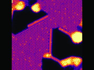
An Oak Ridge National Laboratory–led team has learned how to engineer tiny pores embellished with distinct edge structures inside atomically-thin two-dimensional, or 2D, crystals. The 2D crystals are envisioned as stackable building blocks for ultrathin electronics and other advance...
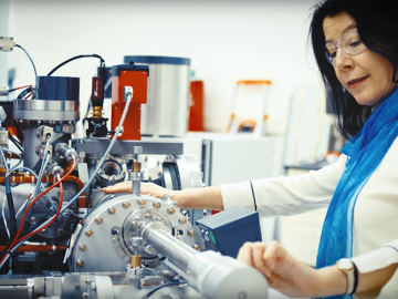
It may take a village to raise a child, according to the old proverb, but it takes an entire team of highly trained scientists and engineers to install and operate a state-of-the-art, exceptionally complex ion microprobe. Just ask Julie Smith, a nuclear security scientist at the Depa...
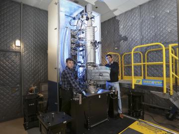
A scientific team led by the Department of Energy’s Oak Ridge National Laboratory has found a new way to take the local temperature of a material from an area about a billionth of a meter wide, or approximately 100,000 times thinner than a human hair. This discove...

Material surfaces and interfaces may appear flat and void of texture to the naked eye, but a view from the nanoscale reveals an intricate tapestry of atomic patterns that control the reactions between the material and its environment. Electron microscopy allows researchers to probe...


