![Man in blue button down shirt poses outside for a picture with his arms crossed.](/sites/default/files/styles/featured_square_large/public/2024-07/Troy_Carter_headshot.jpeg?h=8a7fc05e&itok=VFmZIzHo)
Filter News
Area of Research
News Type
News Topics
- (-) Microscopy (36)
- 3-D Printing/Advanced Manufacturing (82)
- Advanced Reactors (18)
- Artificial Intelligence (75)
- Big Data (31)
- Bioenergy (74)
- Biology (80)
- Biomedical (45)
- Biotechnology (18)
- Buildings (31)
- Chemical Sciences (52)
- Clean Water (15)
- Climate Change (71)
- Composites (15)
- Computer Science (140)
- Coronavirus (34)
- Critical Materials (13)
- Cybersecurity (31)
- Decarbonization (64)
- Education (4)
- Element Discovery (1)
- Emergency (2)
- Energy Storage (70)
- Environment (138)
- Exascale Computing (34)
- Fossil Energy (5)
- Frontier (38)
- Fusion (44)
- Grid (38)
- High-Performance Computing (70)
- Hydropower (5)
- Isotopes (45)
- ITER (4)
- Machine Learning (35)
- Materials (100)
- Materials Science (94)
- Mathematics (6)
- Mercury (9)
- Microelectronics (3)
- Molten Salt (3)
- Nanotechnology (42)
- National Security (54)
- Net Zero (11)
- Neutron Science (96)
- Nuclear Energy (81)
- Partnerships (43)
- Physics (52)
- Polymers (20)
- Quantum Computing (29)
- Quantum Science (56)
- Renewable Energy (2)
- Security (22)
- Simulation (39)
- Software (1)
- Space Exploration (15)
- Statistics (2)
- Summit (50)
- Sustainable Energy (74)
- Transformational Challenge Reactor (7)
- Transportation (52)
Media Contacts
![Starch granules](/sites/default/files/styles/list_page_thumbnail/public/2020-02/starchgranules.png?h=0c9ab501&itok=eLsE3JOx)
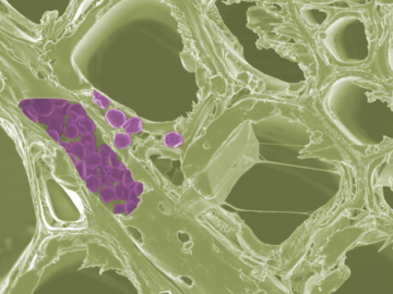
Scientists at the Department of Energy’s Oak Ridge National Laboratory have developed a new method to peer deep into the nanostructure of biomaterials without damaging the sample. This novel technique can confirm structural features in starch, a carbohydrate important in biofuel production.
![Scanning probe microscopes use an atom-sharp tip—only a few nanometers thick—to image materials on a nanometer length scale. The probe tip, invisible to the eye, is attached to a cantilever (pictured) that moves across material surfaces like the tone arm on a record player. Credit: Genevieve Martin/Oak Ridge National Laboratory; U.S. Dept. of Energy.](/sites/default/files/styles/list_page_thumbnail/public/2020-01/2019-P15115.jpg?h=c6980913&itok=o69jyoNw)
Liam Collins was drawn to study physics to understand “hidden things” and honed his expertise in microscopy so that he could bring them to light.
![ORNL alanine_graphic.jpg ORNL alanine_graphic.jpg](/sites/default/files/styles/list_page_thumbnail/public/ORNL%20alanine_graphic.jpg?itok=iRLfcOw-)
OAK RIDGE, Tenn., Jan. 31, 2019—A new electron microscopy technique that detects the subtle changes in the weight of proteins at the nanoscale—while keeping the sample intact—could open a new pathway for deeper, more comprehensive studies of the basic building blocks of life.
![Schematic drawing of the boron nitride cell. Credit: University of Illinois at Chicago. Schematic drawing of the boron nitride cell. Credit: University of Illinois at Chicago.](/sites/default/files/styles/list_page_thumbnail/public/news/images/schematic1.jpg?itok=iYCttAg3)
A new microscopy technique developed at the University of Illinois at Chicago allows researchers to visualize liquids at the nanoscale level — about 10 times more resolution than with traditional transmission electron microscopy — for the first time. By trapping minute amounts of...
![From left, Andrew Lupini and Juan Carlos Idrobo use ORNL’s new monochromated, aberration-corrected scanning transmission electron microscope, a Nion HERMES to take the temperatures of materials at the nanoscale. Image credit: Oak Ridge National Laboratory From left, Andrew Lupini and Juan Carlos Idrobo use ORNL’s new monochromated, aberration-corrected scanning transmission electron microscope, a Nion HERMES to take the temperatures of materials at the nanoscale. Image credit: Oak Ridge National Laboratory](/sites/default/files/styles/list_page_thumbnail/public/news/images/2018-P00413.jpg?itok=UKejk7r2)
A scientific team led by the Department of Energy’s Oak Ridge National Laboratory has found a new way to take the local temperature of a material from an area about a billionth of a meter wide, or approximately 100,000 times thinner than a human hair. This discove...
![ORNL’s Xiahan Sang unambiguously resolved the atomic structure of MXene, a 2D material promising for energy storage, catalysis and electronic conductivity. Image credit: Oak Ridge National Laboratory, U.S. Dept. of Energy; photographer Carlos Jones ORNL’s Xiahan Sang unambiguously resolved the atomic structure of MXene, a 2D material promising for energy storage, catalysis and electronic conductivity. Image credit: Oak Ridge National Laboratory, U.S. Dept. of Energy; photographer Carlos Jones](/sites/default/files/styles/list_page_thumbnail/public/Sang_2016-P07680_0.jpg?itok=w0e5eR_U)
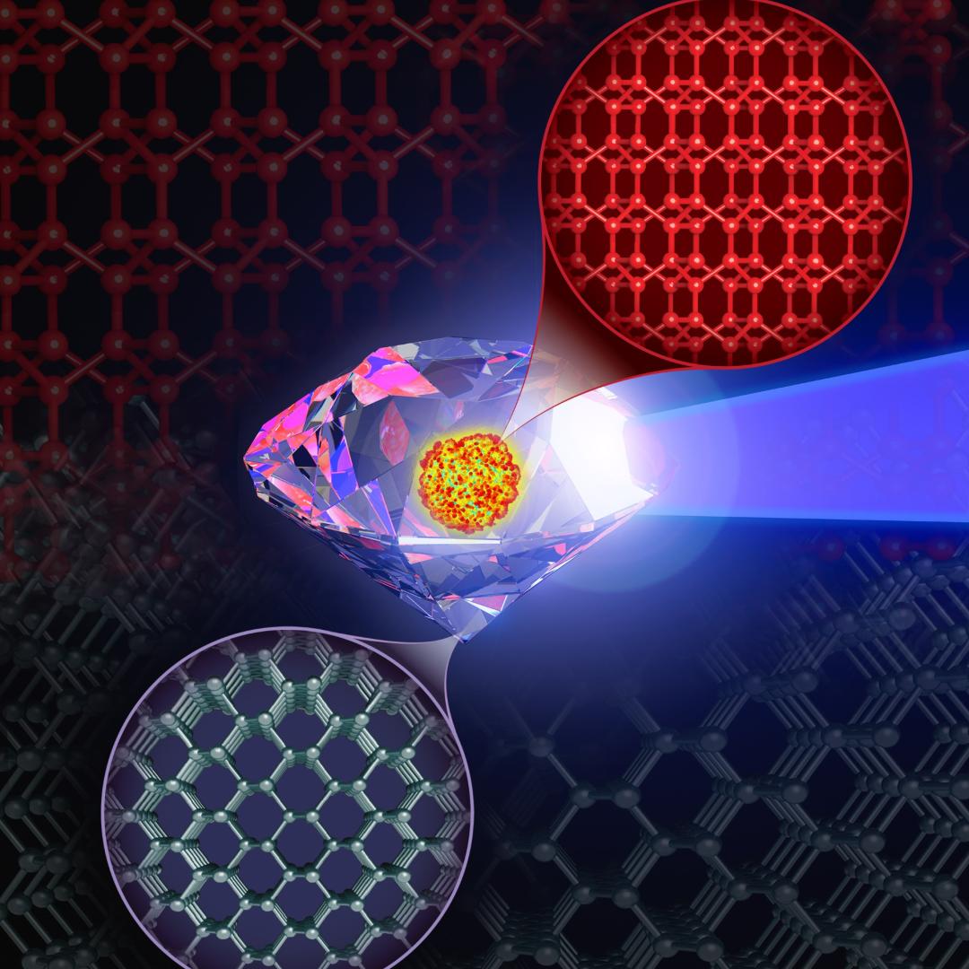
Researchers have long sought electrically conductive materials for economical energy-storage devices. Two-dimensional (2D) ceramics called MXenes are contenders. Unlike most 2D ceramics, MXenes have inherently good conductivity because they are molecular sheets made from the carbides ...