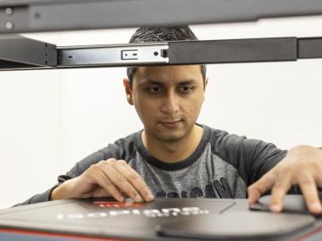
![Sphere that has the top right fourth removed (exposed) Colors from left are orange, dark blue with orange dots, light blue with horizontal lines, then black. Inside the exposure is green and black with boxes.](/sites/default/files/styles/featured_square_large/public/2024-06/slicer.jpg?h=56311bf6&itok=bCZz09pJ)
Filter News
Area of Research
News Topics
- (-) Frontier (1)
- (-) Fusion (13)
- (-) Materials (2)
- (-) Microscopy (8)
- (-) Neutron Science (30)
- (-) Polymers (7)
- 3-D Printing/Advanced Manufacturing (28)
- Advanced Reactors (14)
- Artificial Intelligence (8)
- Big Data (11)
- Bioenergy (12)
- Biology (5)
- Biomedical (21)
- Biotechnology (2)
- Buildings (1)
- Chemical Sciences (5)
- Clean Water (2)
- Climate Change (10)
- Composites (1)
- Computer Science (39)
- Coronavirus (23)
- Critical Materials (2)
- Cybersecurity (4)
- Decarbonization (1)
- Energy Storage (21)
- Environment (29)
- Exascale Computing (3)
- Grid (7)
- High-Performance Computing (3)
- Isotopes (8)
- Machine Learning (8)
- Materials Science (37)
- Mathematics (2)
- Mercury (1)
- Molten Salt (2)
- Nanotechnology (17)
- National Security (2)
- Nuclear Energy (31)
- Physics (13)
- Quantum Science (14)
- Security (3)
- Space Exploration (2)
- Summit (17)
- Sustainable Energy (24)
- Transformational Challenge Reactor (5)
- Transportation (15)
Media Contacts
![Nuclear — Seeing inside particles](/sites/default/files/styles/list_page_thumbnail/public/2020-04/Kernels-nuclear%20materials-2_0.jpg?h=ae51ec69&itok=_AWiopZz)
Oak Ridge National Laboratory researchers working on neutron imaging capabilities for nuclear materials have developed a process for seeing the inside of uranium particles – without cutting them open.
![Postdoctoral researcher Nischal Kafle positions a component for a portable plasma imaging diagnostic device at ORNL in February. The device, a project for ARPA-E, is built of off-the-shelf parts. Credit: Carlos Jones/ORNL](/sites/default/files/styles/list_page_thumbnail/public/2020-03/2020-P00808.jpg?h=8f9cfe54&itok=TGI-lQiS)
The techniques Theodore Biewer and his colleagues are using to measure whether plasma has the right conditions to create fusion have been around awhile.
![Scientists created a novel polymer that is as effective as natural proteins in transporting protons through a membrane. Credit: ORNL/Jill Hemman](/sites/default/files/styles/list_page_thumbnail/public/2020-03/19-G01195_nature_feature_0.png?h=e4fbc3eb&itok=K8czXmTr)
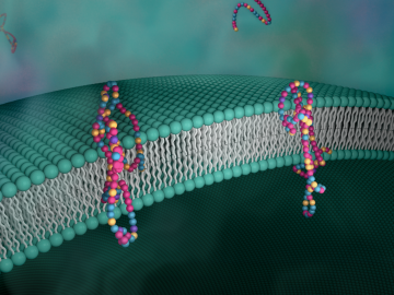
Biological membranes, such as the “walls” of most types of living cells, primarily consist of a double layer of lipids, or “lipid bilayer,” that forms the structure, and a variety of embedded and attached proteins with highly specialized functions, including proteins that rapidly and selectively transport ions and molecules in and out of the cell.
![Polymer self-assembly at the liquid-liquid interface in real time](/sites/default/files/styles/list_page_thumbnail/public/2020-02/descent.png?h=d1cb525d&itok=rz3eSM-H)
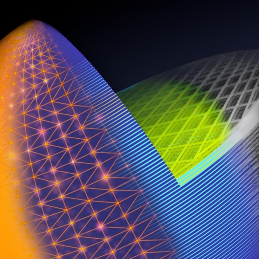
OAK RIDGE, Tenn., Feb. 27, 2020 — Researchers at Oak Ridge National Laboratory and the University of Tennessee achieved a rare look at the inner workings of polymer self-assembly at an oil-water interface to advance materials for neuromorphic computing and bio-inspired technologies.
![Starch granules](/sites/default/files/styles/list_page_thumbnail/public/2020-02/starchgranules.png?h=0c9ab501&itok=eLsE3JOx)
Scientists at the Department of Energy’s Oak Ridge National Laboratory have developed a new method to peer deep into the nanostructure of biomaterials without damaging the sample. This novel technique can confirm structural features in starch, a carbohydrate important in biofuel production.
![This simulation of a fusion plasma calculation result shows the interaction of two counter-streaming beams of super-heated gas. Credit: David L. Green/Oak Ridge National Laboratory, U.S. Dept. of Energy](/sites/default/files/styles/list_page_thumbnail/public/2020-02/Fusion_plasma_simulation.jpg?h=d0852d1e&itok=CDWgjLPL)
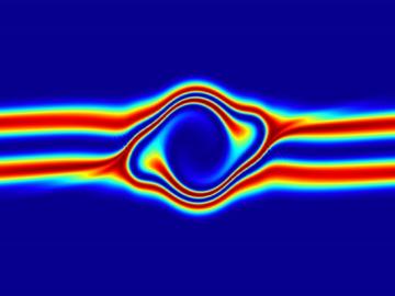
The prospect of simulating a fusion plasma is a step closer to reality thanks to a new computational tool developed by scientists in fusion physics, computer science and mathematics at ORNL.
![Scanning probe microscopes use an atom-sharp tip—only a few nanometers thick—to image materials on a nanometer length scale. The probe tip, invisible to the eye, is attached to a cantilever (pictured) that moves across material surfaces like the tone arm on a record player. Credit: Genevieve Martin/Oak Ridge National Laboratory; U.S. Dept. of Energy.](/sites/default/files/styles/list_page_thumbnail/public/2020-01/2019-P15115.jpg?h=c6980913&itok=o69jyoNw)
Liam Collins was drawn to study physics to understand “hidden things” and honed his expertise in microscopy so that he could bring them to light.